Cell Meter 线粒体膜电位近红外检测试剂盒 适合流式细胞检测
| Ex (nm) | 640 | Em (nm) | 657 |
| 分子量 | - | 溶剂 | - |
| 存储条件 | 在零下15度以下保存, 避免光照 |
产品货期
咨询
产品优势
1.高特异性:依赖于线粒体膜电位(ΔΨm)选择性积聚在活性线粒体中
2.细胞兼容性:适用于增殖细胞、非增殖细胞,兼容悬浮细胞和贴壁细胞的检测。
3.稳定性好:抗光漂白能力强,适合长时间活细胞成像
4.高信噪比:和其他染料相比,信噪比更高,成像更清晰
适用范围
用于标记活细胞
产品介绍
Cell Meter™ 近红外膜电位检测试剂盒是一套通过监测线粒体膜电位(ΔΨm)变化来检测细胞凋亡的专业试剂盒。当细胞凋亡发生时,线粒体膜电位崩溃会引发通透性转换孔(mPTP)开放,导致细胞色素C释放至胞质,进而激活下游凋亡级联反应。
本试剂盒采用专利阳离子染料MitoLite NIR™,在正常细胞中可特异性积聚于线粒体(强荧光信号),而在凋亡细胞中因膜电位丧失导致荧光强度显著降低。该检测兼容流式细胞术(FL4通道)和荧光显微成像,并可与其他试剂联用(如碘化丙啶和Cell Meter™磷脂酰丝氨酸凋亡检测试剂盒),实现细胞活力与凋亡的多参数分析。经优化后适用于通过流式细胞术高通量筛选凋亡激活剂或抑制剂。
适用仪器
| 流式细胞仪 | |
| Ex: | 640 nm |
| Em: | 660/20 nm |
| 通道: | APC 通道 |
样品实验方案
简要概述
1.将待测化合物处理的细胞调整至5×10⁵~1×10⁶ cells/mL密度
2.每1 mL细胞悬液加入5 μL 200× MitoLite™ NIR母液,轻轻涡旋混匀
3. 37℃、5% CO₂培养箱中避光孵育15-30分钟
4.离心细胞,并将细胞重悬于1 mL生长培养基中
5.使用流式细胞仪通过FL4通道(激发635nm,发射670nm)分析细胞
注:本方案适用于大多数哺乳动物细胞系,具体条件需根据细胞类型优化
操作步骤
1.使用预温培养基或自选缓冲液将细胞重悬至5×10⁵~1×10⁶ cells/mL。
注:最佳密度需根据细胞系单独优化
2.加入待测化合物诱导细胞凋亡,并建立平行对照实验。
阴性对照:仅加入化合物溶剂
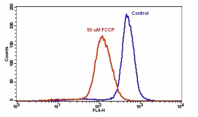
阳性对照:将细胞置于 37 °C、5% CO2 培养箱中,用 5-50 µM FCCP 或 CCCP 处理 15 至 30 分钟。
注意:CCCP 或 FCCP 可与 MitoLite™ NIR 同时添加。建议通过预实验确定各细胞系合适的浓度
3.将 5 µL 200X MitoLite™ NIR(组分 A)添加到处理过的细胞中。
4.将细胞置于 37 °C、5% CO2 培养箱中孵育 15 至 30 分钟。
注意:对于贴壁细胞,用 0.5 mM EDTA 轻轻提起细胞以保持细胞完整,并在与 MitoLite™ NIR 染料上样溶液孵育前,用含血清的培养基清洗一次。
5. 1000 rpm离心4分钟,使用1 mL Assay Buffer(组分B)或自选缓冲液重悬细胞。
6.使用流式细胞仪通过FL4通道(激发635nm,发射670nm)分析细胞。