Portelite 荧光法DNA 定量试剂盒*检测限更广* *适用于 Cytocite 和 Qubit 荧光仪*
| Ex (nm) | - | Em (nm) | - |
| 分子量 | - | 溶剂 | - |
| 存储条件 | - |
在进行各种分析的DNA样品制备中,DNA定量是一项非常重要的任务。 Portelite 荧光DNA定量试剂盒提供了使用Qubit荧光仪操作Helixyte Green BR快速定量dsDNA的方法。它针对Cytocite 和Qubit 荧光仪进行了优化。 Portelite 荧光DNA定量测定在三个数量级上呈线性。该测定法对RNA上的双链DNA(dsDNA)具有高度选择性,并针对测量10 pg / µL至10 ng / µL的dsDNA浓度进行了优化。Helixyte Green-BR与dsDNA结合后表现出较大的荧光增强,并且比UV吸光度读数高几个数量级。百萤生物是AAT Bioquest的中国代理商,为您提供优质的Helixyte 绿色双链DNA定量检测试剂盒。
适用仪器
| 量子位荧光计 | |
| Ex: | 480 nm |
| Em: | 530 nm |
| 规格: | 0.2 mL PCR 小瓶 |
|
CytoCite 荧光计 |
|
| Ex: | 480 nm |
| Em: | 530 nm |
| 规格: | 0.2 mL PCR小瓶 |
样品实验方案
简要概述
- 准备Helixyte Green BR工作溶液
- 在每个0.2 mL PCR管中加入190 uL 1X Helixyte Green BR工作溶液
- 在每个试管中添加10 uL DNA标准溶液或测试样品
- 在室温下孵育2分钟
- 使用CytoCite 荧光仪或Qubit 检测荧光
注意:打开之前,请将所有成分置于室温下。
溶液配制
工作溶液配制
Helixyte Green-BR工作溶液:在DNA分析缓冲液(组分B)中稀释200倍Portelite dsDNA试剂(组分A)。 例如,要为8个样品准备足够的工作溶液,请将5uL Helixyte Green BR(组分A)添加到1mL DNA分析缓冲液(组分B)中。注意:通过用箔纸覆盖或将其置于黑暗中来保护工作液免受光照。 我们建议在塑料容器中操作而不是玻璃中制备该溶液,因为染料可能会吸附到玻璃表面。 为了获得佳结果,应在准备后的几个小时内使用此溶液。
实验步骤
根据DNA样品的估计浓度,样品量可以在1〜20 uL的范围内。推荐的样品量为10 uL,DNA浓度在0.2〜100 ng / uL范围内。如果使用其他样品体积,请在浓度计算中调整稀释倍数。
10 uL样品体积(DNA浓度在0.2〜100 ng / uL范围内)实验方案。
- 在每个Cytocite 样品管(#CCT100)或等效的0.2 mL PCR管中添加190 uL 1X Helixyte Green BR工作溶液。注意:请使用薄壁聚丙烯透明0.2 mL PCR管,例如#CCT100。
- 向每个试管中加入10 µL DNA标准品或测试样品,然后离心2〜3秒进行混合。
- 将所有试管在室温下孵育2分钟。
- 将样品插入CytoCite 或Quibit 中,并通过绿色荧光通道检测荧光。若使用CytoCite,请遵循适用于CytoCite 荧光仪的程序。
标准校准曲线的准备
对于Portelite 分析,您可以选择使用DNA标准液绘制校正曲线。
- 在DNA分析缓冲液(成分B)中,用100 ng/uL的DNA标准BR#2(成分D)进行1/3系列稀释,得到30、10、3、1、0.3、0.1和0 ng/uL的DNA标准稀释液。
- 在每个试管中加入190 uL Helixyte Green BR工作溶液。
- 将10 uL标准液或10 uL样品添加到0.2 mL PCR管中。
- 将反应在室温下孵育2分钟。
- 将样品插入CytoCite ,并通过绿色荧光通道检测荧光。
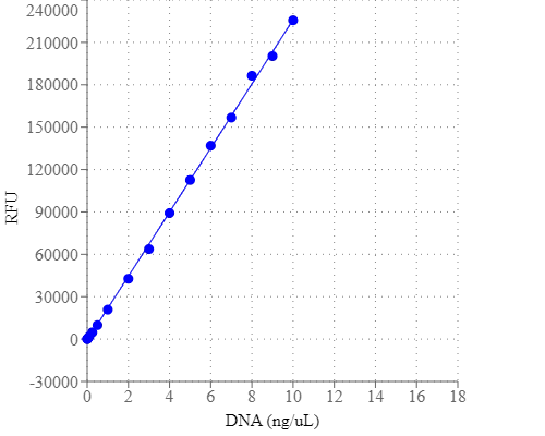
图示
 图1.使用具有宽动态范围的Portelite 荧光DNA定量试剂盒生成的DNA标准曲线。 使用绿色荧光通道定量荧光强度,使用线性拟合计算回归模型。 |
参考文献
A new reporter design based on DNA origami nanostructures for quantification of short oligonucleotides using microbeads
Authors: Choi, Y., Schmidt, C., Tinnefeld, P., Bald, I., Rodiger, S.
Journal: Sci Rep (2019): 4769
A universal fluorescence-based toolkit for real-time quantification of DNA and RNA nuclease activity
Authors: Sheppard, E. C., Rogers, S., Harmer, N. J., Chahwan, R.
Journal: Sci Rep (2019): 8853
Effects of Quantification Methods, Isolation Kits, Plasma Biobanking, and Hemolysis on Cell-Free DNA Analysis in Plasma
Authors: Streleckiene, G., Forster, M., Inciuraite, R., Lukosevicius, R., Skieceviciene, J.
Journal: Biopreserv Biobank (2019): ersion="1.0" encoding="UTF-8" ?>17645.enlEndNote1117Streleckiene, G.Forster, M.Inciuraite, R.Lukosevicius, R.Skieceviciene, J.1Institute for Digestive Research, Lithuanian University of Health Sciences, Kaunas, Lithuania. 2Institute of Clinical Molecu
Flat-top TIRF illumination boosts DNA-PAINT imaging and quantification
Authors: Stehr, F., Stein, J., Schueder, F., Schwille, P., Jungmann, R.
Journal: Nat Commun (2019): 1268
Molecular-Recognition-Based DNA Nanodevices for Enhancing the Direct Visualization and Quantification of Single Vesicles of Tumor Exosomes in Plasma Microsamples
Authors: He, D., Ho, S. L., Chan, H. N., Wang, H., Hai, L., He, X., Wang, K., Li, H. W.
Journal: Anal Chem (2019): 2768-2775
Quantification of fixed adherent cells using a strong enhancer of the fluorescence of DNA dyes
Authors: Ligasova, A., Koberna, K.
Journal: Sci Rep (2019): 8701
A fluorescent reporter for quantification and enrichment of DNA editing by APOBEC-Cas9 or cleavage by Cas9 in living cells
Authors: St Martin, A., Salamango, D., Serebrenik, A., Shaban, N., Brown, W. L., Donati, F., Munagala, U., Conticello, S. G., Harris, R. S.
Journal: Nucleic Acids Res (2018): e84
Accuracy of human sperm DNA oxidation quantification and threshold determination using an 8-OHdG immuno-detection assay
Authors: Vorilhon, S., Brugnon, F., Kocer, A., Dollet, S., Bourgne, C., Berger, M., Janny, L., Pereira, B., Aitken, R. J., Moazamian, A., Gharagozloo, P., Drevet, J., Pons-Rejraji, H.
Journal: Hum Reprod (2018): 553-562
Cell Type-Specific Quantification of Telomere Length and DNA Double-strand Breaks in Individual Lung Cells by Fluorescence In Situ Hybridization and Fluorescent Immunohistochemistry
Authors: van Batenburg, A. A., Kazemier, K. M., Peeters, T., van Oosterhout, M. F. M., van der Vis, J. J., Grutters, J. C., Goldschmeding, R., van Moorsel, C. H. M.
Journal: J Histochem Cytochem (2018): 485-495
Identification and Quantification of Heterogeneously-methylated DNA Fragments Using Epiallele-sensitive Droplet Digital Polymerase Chain Reaction (EAST-ddPCR)
Authors: Menschikowski, M., J and eck, C., Friedemann, M., Richter, S., Thiem, D., Lange, B. S., Suttorp, M.
Journal: Cancer Genomics Proteomics (2018): 299-312