Cell Navigator 细胞膜染色试剂盒
英文名称:Cell Navigator® Cell Plasma Membrane Staining Kit *Green Fluorescence*
产品参数
| Ex (nm) | 497 | Em (nm) | 505 |
| 分子量 | - | 溶剂 | - |
| 存储条件 | 在零下15度以下保存, 避免光照 |
产品概述
产品货期
咨询
产品优势
可作为HCS的细胞分割工具,也可用于标准荧光显微镜下的细胞膜染色
适用范围
用于细胞膜染色
产品介绍
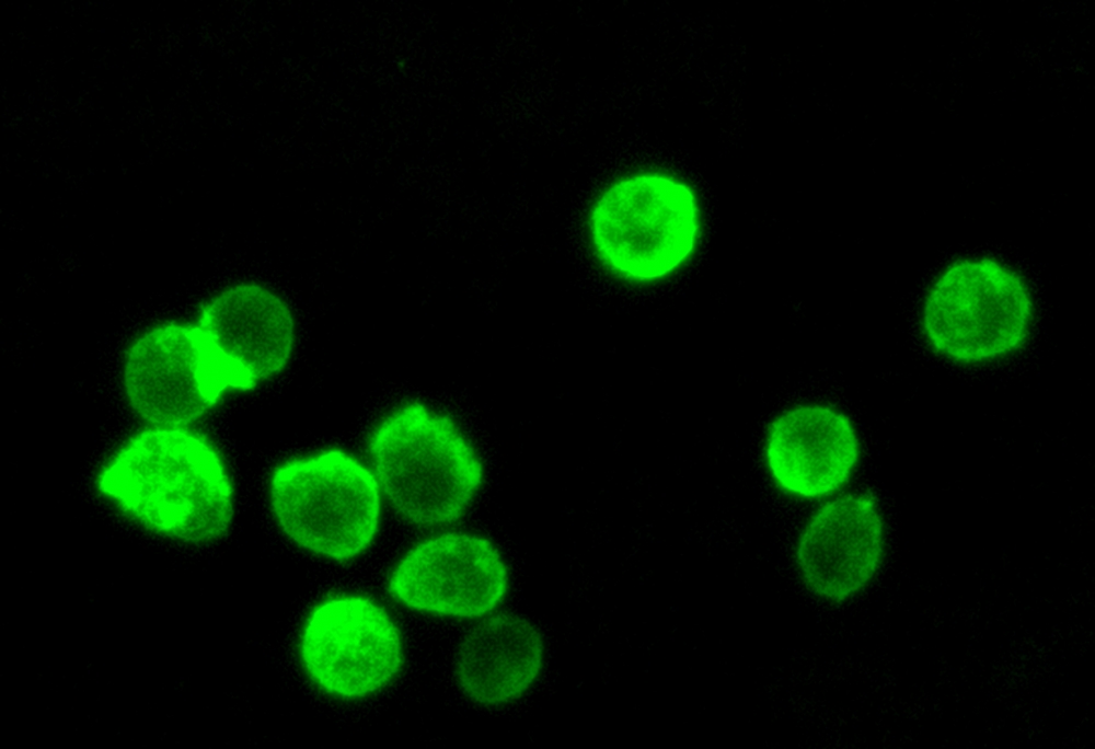
Cell Navigator® 细胞膜染色试剂盒能够快速、均匀地对细胞膜进行标记,且不会出现凝集素染色细胞类型差异。该试剂盒既可作为HCS的细胞分割工具,也可用于标准荧光显微镜下的细胞膜染色。试剂盒所含染料可耐受固定处理,但无法耐受透化操作,因此不适用于需通过抗体检测细胞内靶点的实验。
适用仪器
| 荧光显微镜 | |
| Ex: | FITC 滤波片组 |
| Em: | FITC 滤波片组 |
| 推荐孔板: | 黑色透明底板 |
实验方案
样品实验方案
简要概述
1.在生长培养基中准备细胞
2.准备并向细胞添加Cellpaint Green工作溶液
3.在37℃下孵育5至20分钟
4.使用FITC滤光片组读取荧光强度
溶液配制
储备溶液配制
所有未使用的储备溶液应分为一次性使用的等分试样,并在制备后储存在-20°C下。 避免重复冻融。
Cellpaint 绿色原液(500X):
将100 uL DMSO(组分C)添加到Cellpaint Green(组分A)小瓶中,制成500X储备溶液。
注意:20 µL Cellpaint Green 500X储备溶液可满足一个96孔板。 如果管密封严密,可以将未使用的Cellpaint Green 500X储备溶液分装并在≤-20ºC下保存1个月。 避光并避免重复的冻融。
工作溶液配制
Cellpaint 绿色工作溶液(1X):
将20 uL的500X储备溶液添加到10 mL的测定缓冲液(组分B)中,并充分混合。
注意:我们建议现配现用
样品示例及操作
1.在细胞板中加入100 uL /孔(96孔板)或50 uL /孔(384孔板)的Cellpaint Green工作溶液。 避光保存,在37ºC下孵育细胞5-20分钟。
注意:细胞膜探针的浓度取决于具体应用。 可以根据特定的细胞类型和细胞或组织对探针的渗透性来改变染色条件。
2.除去每个孔中的工作溶液。 用生理缓冲液(例如HHBS,DPBS或您选择的缓冲液)洗涤细胞3次,并替换为HHBS。
3.可选:染色后固定细胞。 用4%甲醛固定细胞15-30分钟。 用生理缓冲液洗涤细胞三遍。
4.使用带有FITC滤波片组的荧光显微镜观察细胞中的荧光信号。
2.除去每个孔中的工作溶液。 用生理缓冲液(例如HHBS,DPBS或您选择的缓冲液)洗涤细胞3次,并替换为HHBS。
3.可选:染色后固定细胞。 用4%甲醛固定细胞15-30分钟。 用生理缓冲液洗涤细胞三遍。
4.使用带有FITC滤波片组的荧光显微镜观察细胞中的荧光信号。